Oct 5 • AFK study plan

Essential Differentials from Vesiculobullous Disease to Jaw Cysts

Whether you are preparing for clinical board examinations or refining your diagnostic accuracy in everyday practice, oral pathology requires mastering complex microscopic patterns, genetic mutations, and radiographic signs. Oral tissue often serves as the primary window into systemic health.

🎁 Your FREE AFK High-Yield Guide Is Ready!

Get instant access to a high-yield AFK PDF packed with important concepts, key facts, and exam-focused information you need to know.
📘 Study smart. Focus on what matters. Build your confidence.
Good luck with your AFK journey! 🦷💙
Enter your email to get your FREE PDF instantly.
Write your awesome label here.

1. Vesiculobullous Lesions: Autoimmune vs. Viral Differentiation
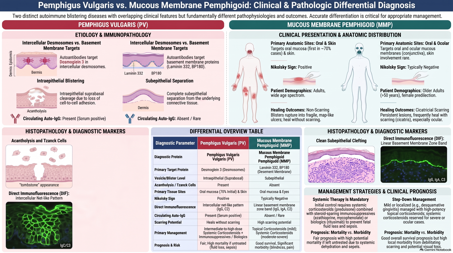
Painful oral blistering that rapidly ruptures into superficial ulcerations requires careful immunologic and histological differentiation.
1- Autoimmune Blistering Diseases
• Pemphigus Vulgaris:
Driven by circulating IgG autoantibodies against Desmoglein 3, targeting epithelial desmosomes. Histology demonstrates suprabasal intraepithelial acantholysis, leaving the basal layer attached to the basement membrane alongside free-floating, rounded Tzanck cells. Application of gentle mechanical friction to normal mucosa yields a positive Nikolsky sign.
• Mucous Membrane Pemphigoid (MMP):
Caused by autoantibodies against basement membrane proteins, specifically laminin 5 and BP180. Histology reveals subepithelial clefting without acantholysis. Direct immunofluorescence (DIF) displays a linear continuous band of IgG and C3 along the basement membrane zone.
 2- Viral Pathologies
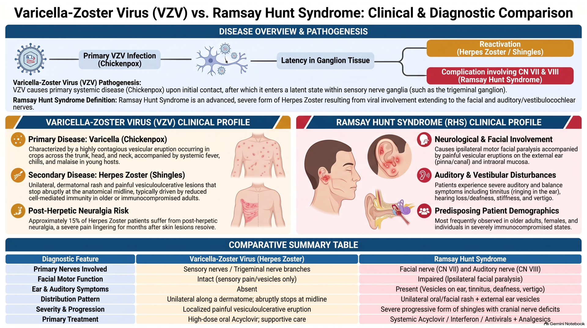
Varicella-Zoster Virus (VZV):
Reactivation of latent VZV in sensory ganglia leads to herpes zoster (shingles), causing painful unilateral vesicular eruptions along dermatomes .
Ramsay Hunt Syndrome:
Occurs when VZV involves the facial and auditory nerves, presenting with ipsilateral facial paralysis, external ear vesicles, tinnitus, vertigo, and deafness.

2. Cysts of the Jaws & Neck: Key Radiographic & Pathological Features
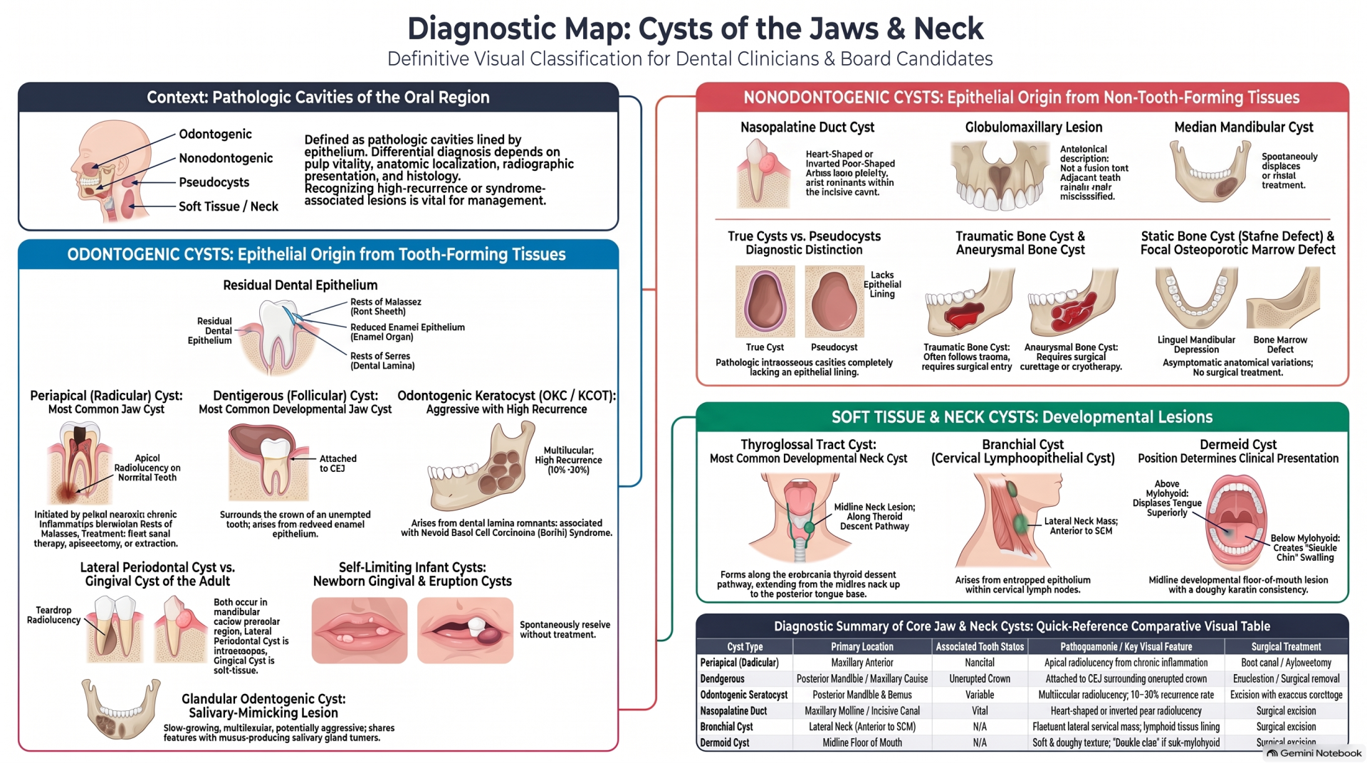
A true cyst is an epithelial-lined pathologic cavity, whereas a pseudocyst lacks an epithelial lining.
1- Radicular (Periapical) Cyst:
The most common inflammatory jaw cyst, arising from rests of Malassez in response to pulpal necrosis in a nonvital tooth.
2- Dentigerous (Follicular) Cyst:
The most common developmental odontogenic cyst, attached precisely at the cementoenamel junction (CEJ) of an unerupted or impacted tooth crown. It originates from the proliferation of the reduced enamel epithelium.
3- Odontogenic Keratocyst (OKC):
Derived from dental lamina remnants (rests of Serres). OKC is known for aggressive clinical behavior, multilocular expansion, and a high rate of recurrence.
4- Nasopalatine Duct Cyst:
The most common non-odontogenic cyst, located within the incisive canal and presenting as a heart-shaped radiolucency at the anterior midline.

Free Resources to Support Your Preparation 🎥📚🆓

We believe that every candidate should have access to high-quality learning materials, which is why we offer free resources to support your preparation for the AFK exam! 🌟🎯📖

Interactive Videos: Our expertly crafted videos explain key topics in an engaging, easy-to-understand format, helping you grasp essential concepts more effectively.

Question Bank with Detailed Answers: Practice with real exam-style questions and receive comprehensive explanations to strengthen your understanding and improve your problem-solving skills.

One-on-One Consultation: Need a study plan tailored to your unique needs? We offer personalized 1-on-1 sessions, where we assess your situation and learning preferences to recommend the best study strategies for your success. 🤝📈📅

These resources are designed to empower you with the knowledge and confidence needed to excel in the equivalency process. Take advantage of them and set yourself up for success! 🚀📚💡

3. Metabolic & Endocrine Bone Disorders
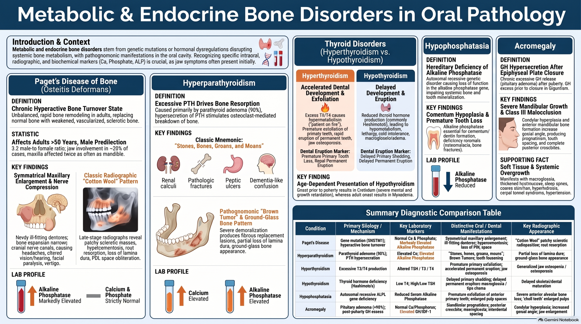
Systemic metabolic disturbances directly impact alveolar bone remodeling, root structure, and jaw density.

4. Hereditary Craniofacial & Developmental Anomalies
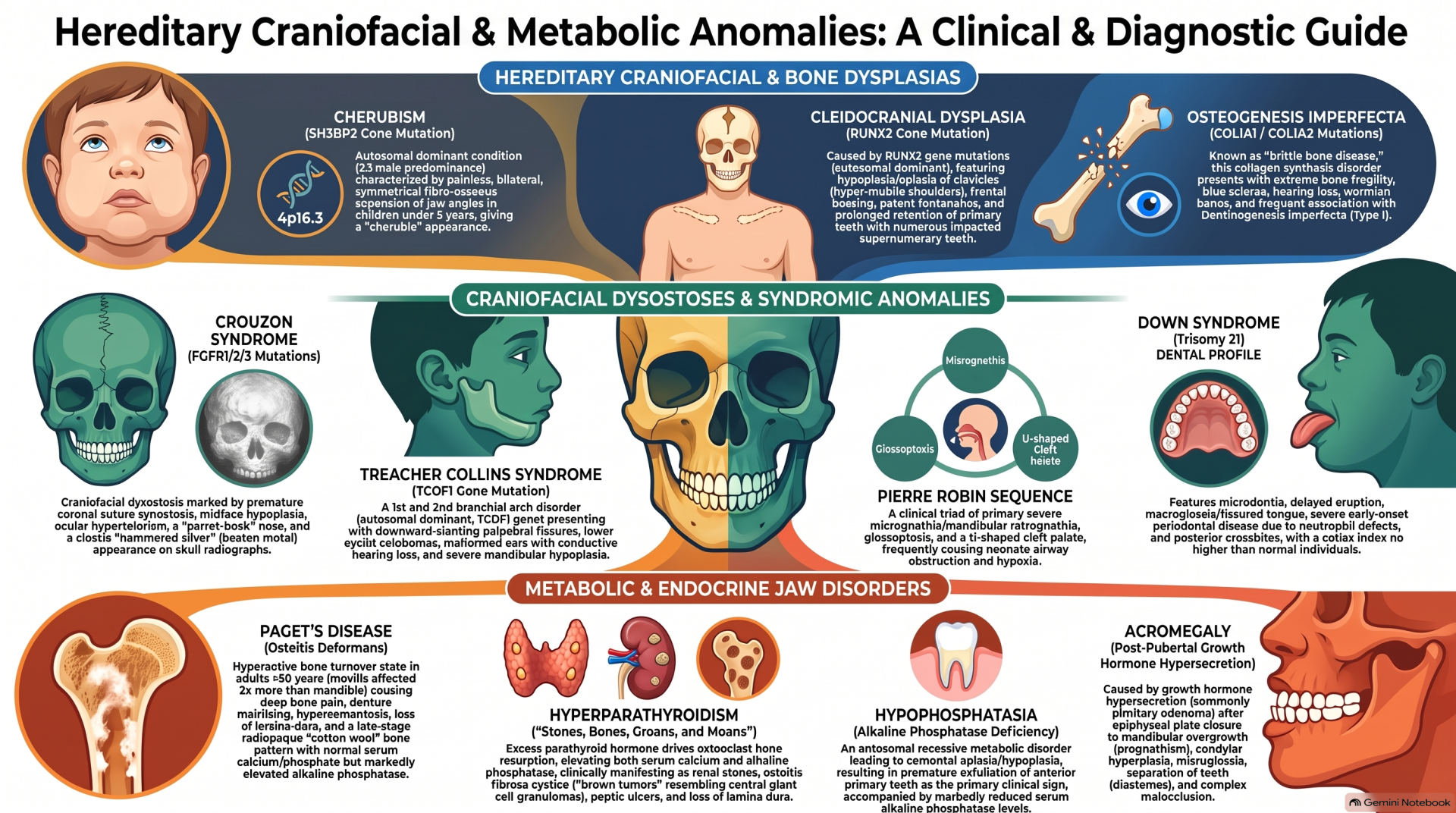
Recognizing inherited genetic mutations prevents diagnostic delay during pediatric clinical evaluation.
1- Cherubism:
An autosomal dominant condition caused by mutations in the SH3BP2 gene (chromosome 4p16.3). It causes painless, bilateral symmetrical enlargement of the posterior mandibles and ascending rami, imparting a characteristic "cherubic" upward-gazing eye appearance in children.
2- Cleidocranial Dysplasia (CCD):
Caused by mutations in the RUNX2 transcription factor. Clinical hallmarks include aplastic or hypoplastic clavicles allowing hypermobility of the shoulders, persistent open fontanelles, maxillary hypoplasia, and numerous unerupted supernumerary teeth.
3- Treacher Collins Syndrome (Mandibulofacial Dysostosis):
Associated with TCOF1 gene mutations, affecting structures derived from the first and second branchial arches. Characterized by downward slanting palpebral fissures, lower eyelid colobomas, malformed ears, and severe zygomatic and mandibular hypoplasia.
4- Pierre Robin Sequence:
Defined by the clinical triad of micrognathia, glossoptosis, and a high-arched or cleft palate, frequently leading to severe neonatal airway obstruction

5. Connective Tissue Lesions: Reactive Hyperplasias vs. Neoplasms
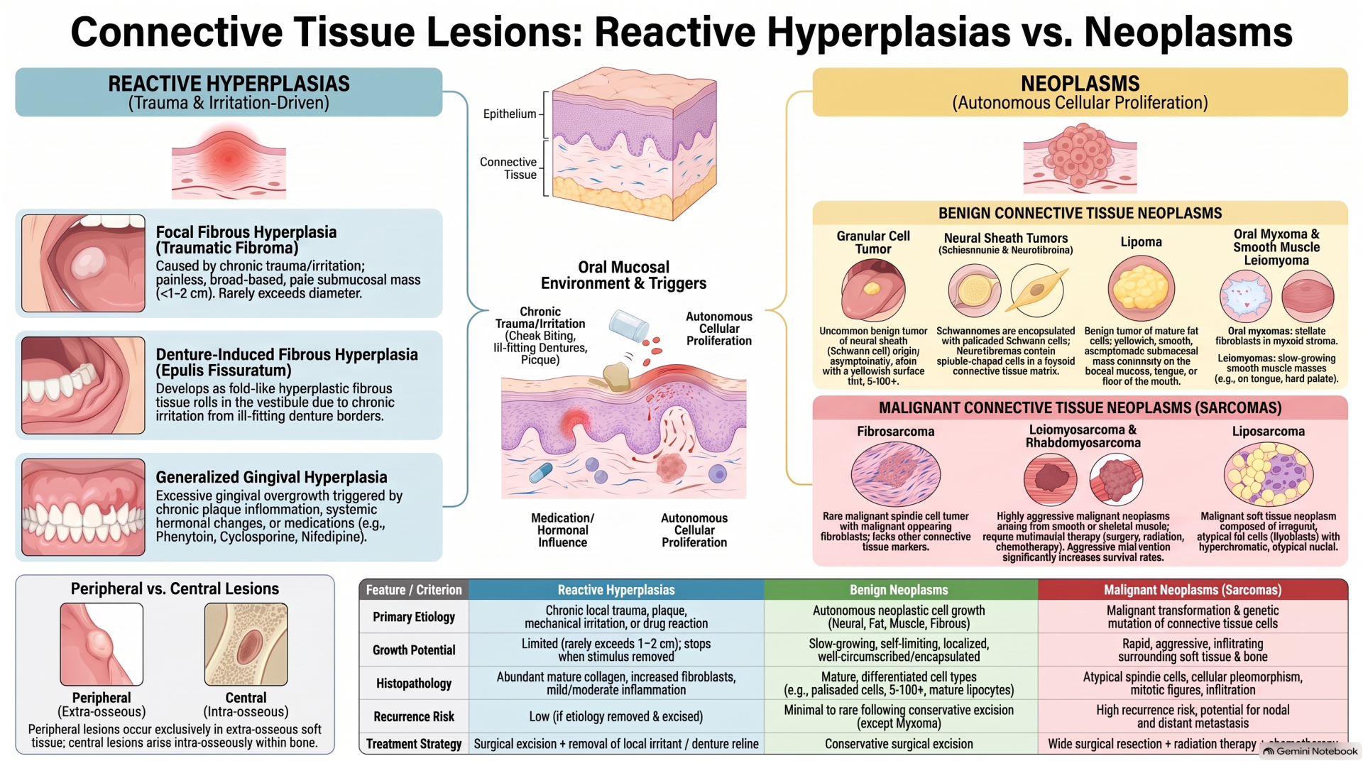
Connective tissue swellings are broadly divided into reactive inflammatory repair responses and true neoplasms.
1- Focal Fibrous Hyperplasia (Irritational Fibroma):
The most common reactive lesion of the oral mucosa, caused by chronic low-grade mechanical trauma (e.g., cheek biting). Histology shows mature fibroblasts scattered within a dense, hyperfunctional collagen matrix.
2- Denture-Induced Fibrous Hyperplasia (Epulis Fissuratum):
Hyperplastic folds of fibrous tissue developing in the alveolar vestibule secondary to chronic irritation from ill-fitting denture flanges.
3- Fibromatosis:
A locally aggressive, non-metastasizing fibrous neoplasm characterized by infiltrative, destructive growth and high recurrence potential

6. Hematologic Manifestations in the Oral Cavity
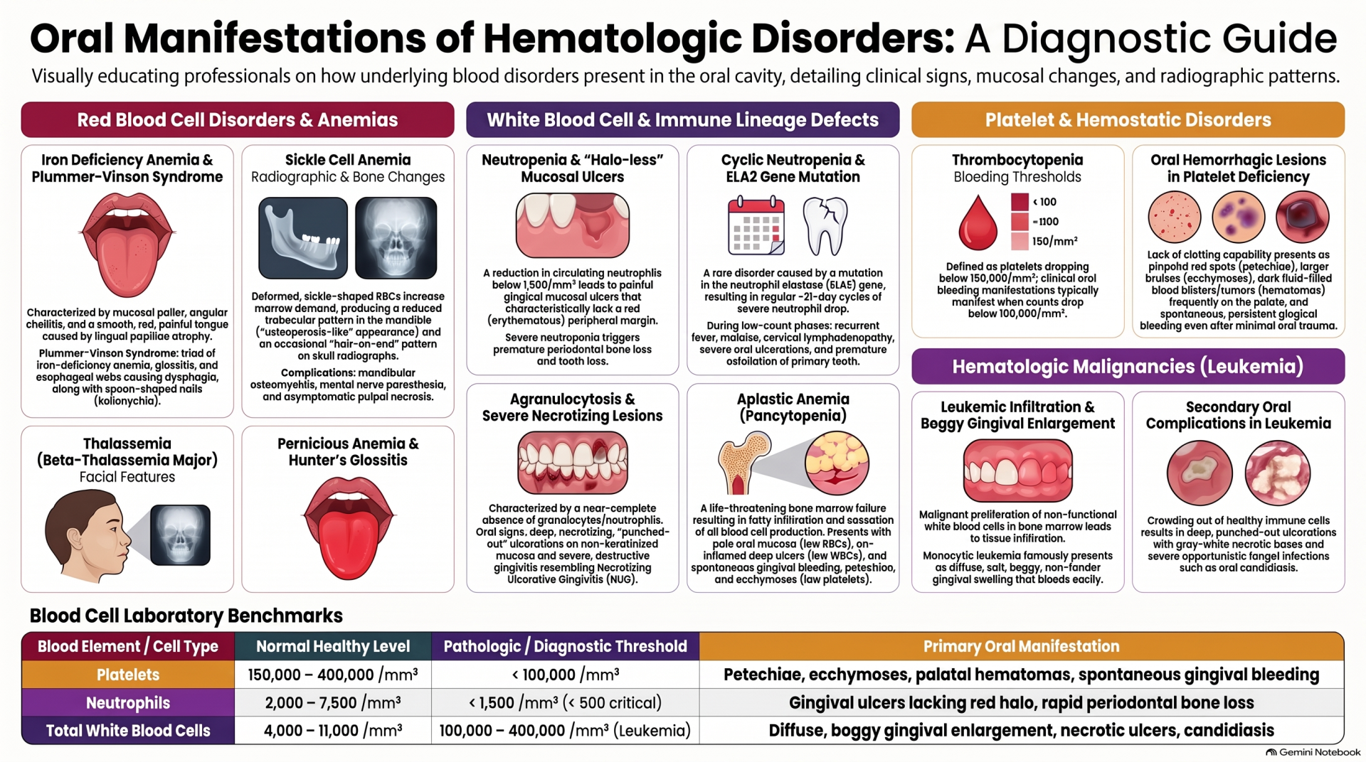
Because the oral mucosa undergoes rapid cellular turnover, systemic blood dyscrasias frequently manifest first in the mouth.
1- Neutropenia:
A drop in circulating neutrophils below 1,500/mm³ triggers deep, painful oral ulcerations that characteristically lack an erythematous halo. Managed using recombinant human G-CSF and targeted antibiotics.
2- Thrombocytopenia:
Platelet reduction below 100,000/mm³ leads to spontaneous mucosal bleeding, petechiae, ecchymoses, and palatal hematomas.
3- Anemic Glossitis:
Severe iron or vitamin deficiencies lead to Hunter's or Moeller's glossitis, characterized by complete atrophy of filiform papillae, resulting in a smooth, red, burning tongue surface

About the Author:

Dr. Mohamed is a licensed dentist in Canada who successfully passed both AFK and ACJ exams. After seeing too many talented international dentists fail due to poor study strategies, he created AFKStudyPlan to provide structured, evidence-based preparation. He's helped 342+ dentists pass their NDEB equivalency exams.
Have questions?
Email us at Info@afkstudyplan.com
or call 587-707-7068.

Latest from our blog