Oct 11 • AFK study plan

The Modern ICCMS™ & ICDAS Framework: Evidence-Based Caries Risk Assessment and Staging

Traditional classification systems like G.V. Black’s (Class I through VI) focus strictly on mechanical cavity preparation after structural tooth damage has already occurred. In contrast, the International Caries Classification and Management System (ICCMS™) and International Caries Detection and Assessment System (ICDAS) provide a holistic, health-outcomes-focused medical framework. By integrating patient-level risk assessment, visual and radiographic lesion staging, and lesion activity assessment, clinicians can deliver personalized, tooth-preserving care.

🎁 Your FREE AFK High-Yield Guide Is Ready!

Get instant access to a high-yield AFK PDF packed with important concepts, key facts, and exam-focused information you need to know.
📘 Study smart. Focus on what matters. Build your confidence.
Good luck with your AFK journey! 🦷💙
Enter your email to get your FREE PDF instantly.
Write your awesome label here.

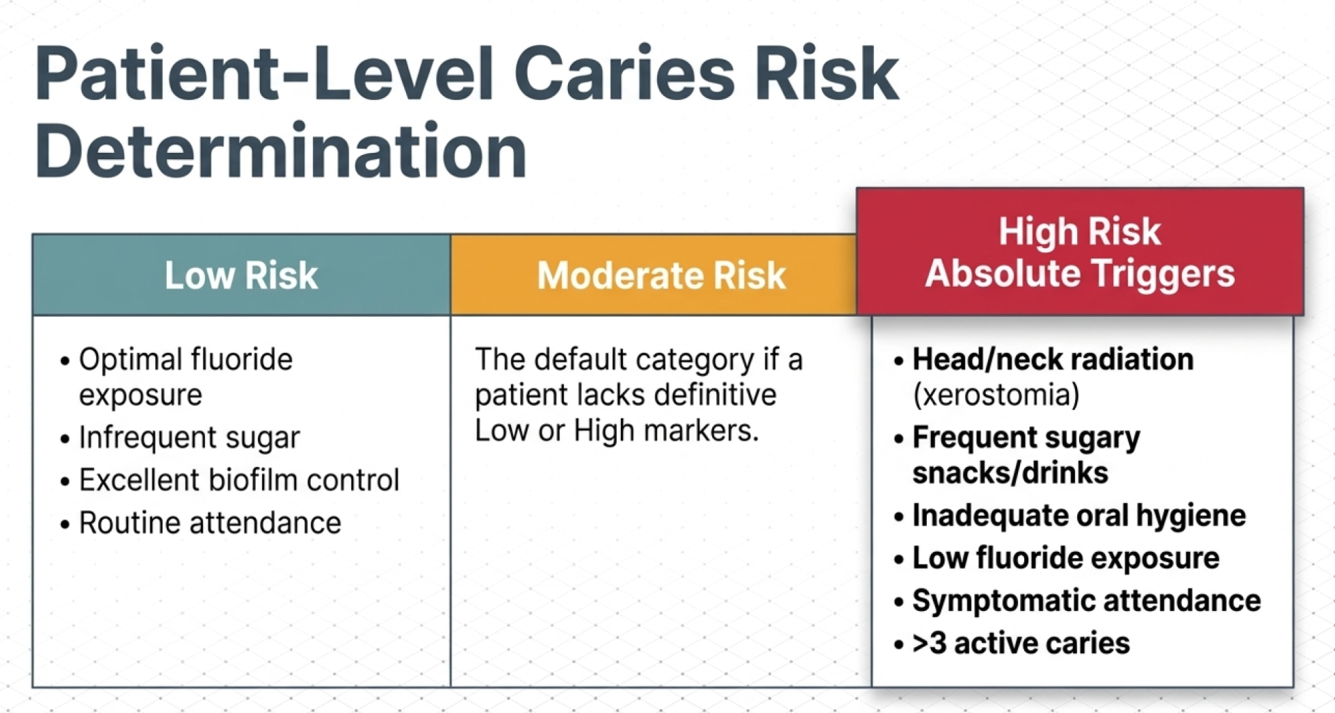
Step 1: Patient-Level & Intraoral Caries Risk Assessment
Step 2: Visual (ICDAS) & Radiographic (R) Staging
Step 2: Visual (ICDAS) & Radiographic (R) Staging
Visual examination requires complete plaque removal and 5 seconds of surface air drying, complemented by bitewing radiographs to evaluate hidden proximal surfaces.
Visual ICDAS Codes & Merged Categories
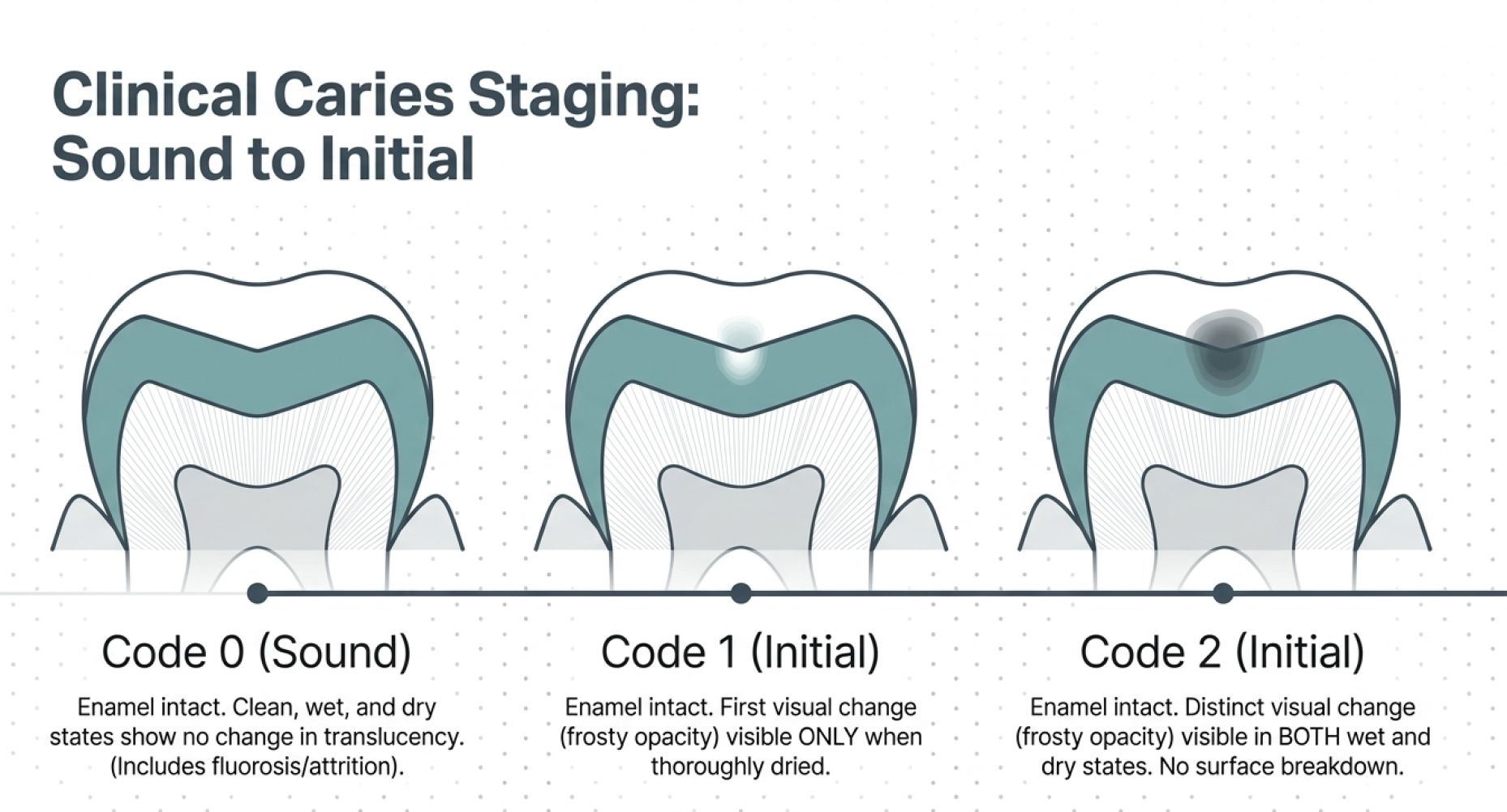
1- Sound Surfaces (ICDAS Code 0): Enamel displays no visual evidence of caries after 5 seconds of air drying. Developmental anomalies, enamel fluorosis, and non-carious tooth wear (attrition, abrasion, erosion) are recorded as sound because the framework evaluates caries pathology exclusively.
2- Initial Stage Caries (ICDAS Codes 1 & 2): Code 1 represents the first visual opacity/discoloration visible only after prolonged air drying. Code 2 displays a distinct white or brown spot lesion visible on both wet and dry enamel, without surface breakdown or underlying dentin shadowing.
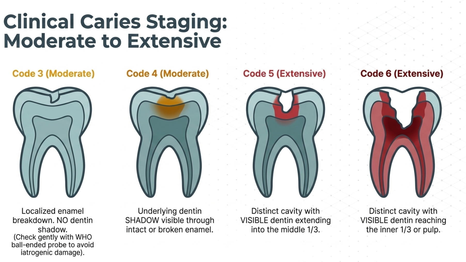
3- Moderate Stage Caries (ICDAS Codes 3 & 4): Code 3 features localized enamel micro-cavitation without exposed dentin. Code 4 manifests as a dark, underlying dentin shadow showing through intact or micro-fractured enamel. Enamel integrity is confirmed gently with a rounded WHO/CPI ball-ended probe to prevent iatrogenic enamel breakdown.
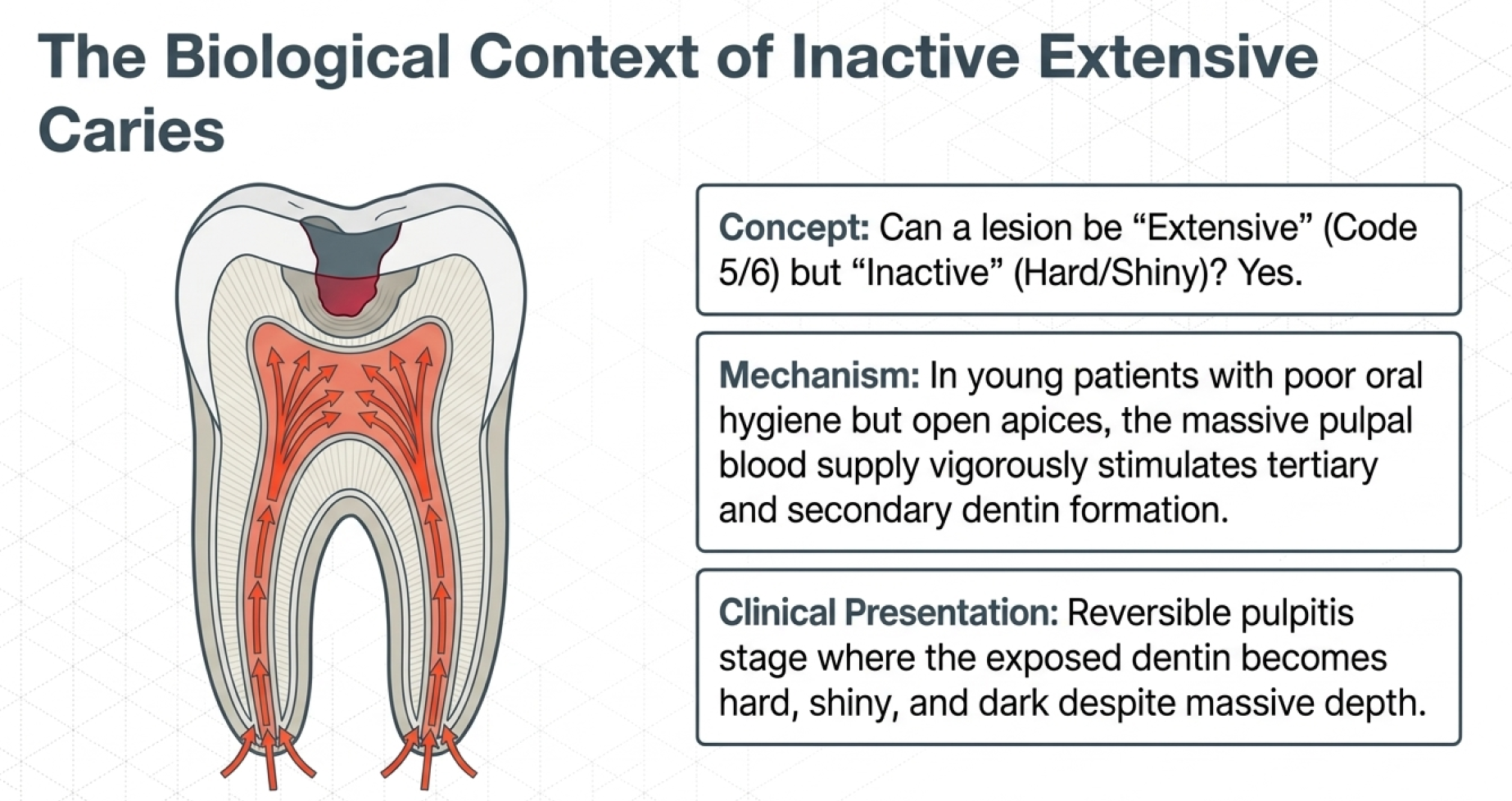
4- Extensive Stage Caries (ICDAS Codes 5 & 6): Code 5 exhibits a distinct cavity with clinically visible dentin extending into the outer or middle third of dentin. Code 6 presents as a deep, extensive cavity involving the inner third of dentin or exposing the pulp
Radiographic Scoring System (R Categories):
1- R0 (Sound):
No radiolucency present.
2- Initial Radiographic Stages (RA1, RA2, RA3):
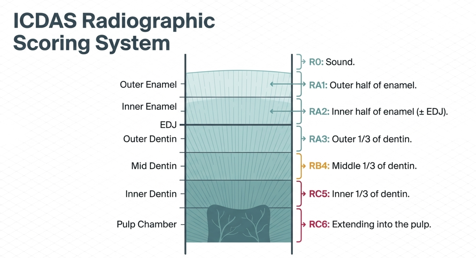
RA1 indicates radiolucency in the outer half of enamel.
RA2 shows radiolucency in the inner half of enamel up to the dentino-enamel junction (DEJ). 
RA3 extends into the outer third of dentin; it remains classified as an Initial lesion because clinical studies demonstrate that only ~32% of outer-third dentinal radiolucencies manifest cavitation.
3- Moderate Radiographic Stage (RB4):
Radiolucency extending into the middle third of dentin.
4- Extensive Radiographic Stages (RC5, RC6):
RC5 extends into the inner third of dentin with clinical cavitation, while RC6 penetrates directly into the pulp.

Free Resources to Support Your Preparation 🎥📚🆓

We believe that every candidate should have access to high-quality learning materials, which is why we offer free resources to support your preparation for the AFK exam! 🌟🎯📖

Interactive Videos: Our expertly crafted videos explain key topics in an engaging, easy-to-understand format, helping you grasp essential concepts more effectively.

Question Bank with Detailed Answers: Practice with real exam-style questions and receive comprehensive explanations to strengthen your understanding and improve your problem-solving skills.

One-on-One Consultation: Need a study plan tailored to your unique needs? We offer personalized 1-on-1 sessions, where we assess your situation and learning preferences to recommend the best study strategies for your success. 🤝📈📅

These resources are designed to empower you with the knowledge and confidence needed to excel in the equivalency process. Take advantage of them and set yourself up for success! 🚀📚💡

Step 3: Lesion Activity Assessment (Active vs. Inactive)
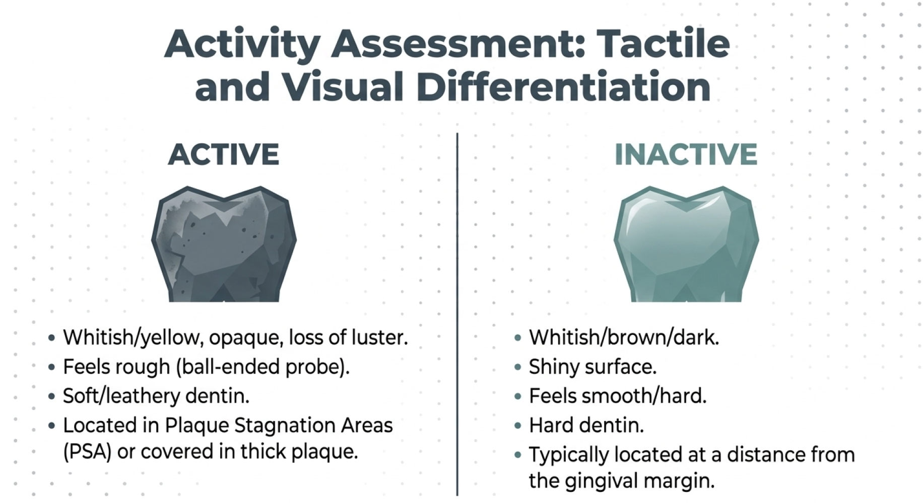
Determining whether a lesion is active or arrested dictates whether therapeutic intervention is required:
1- Active Lesions: Enamel appears opaque, whitish/yellowish, and matte/rough upon gentle tactile probing in plaque stagnation areas (pits, fissures, gingival margins, or below proximal contact points). Active dentin lesions feel soft or leathery.
2- Inactive (Arrested) Lesions: Enamel appears shiny, whitish/brownish/black, and feels smooth and hard upon gentle tactile probing. Inactive dentin lesions feel hard and dark.

Step 4: Risk-Adjusted Clinical Management Pathways
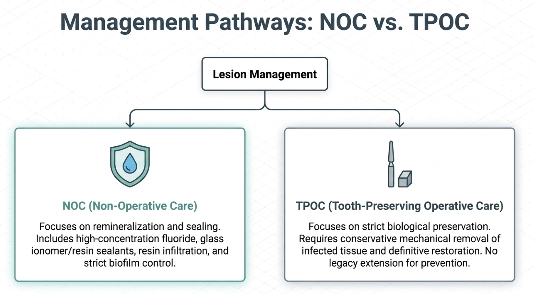
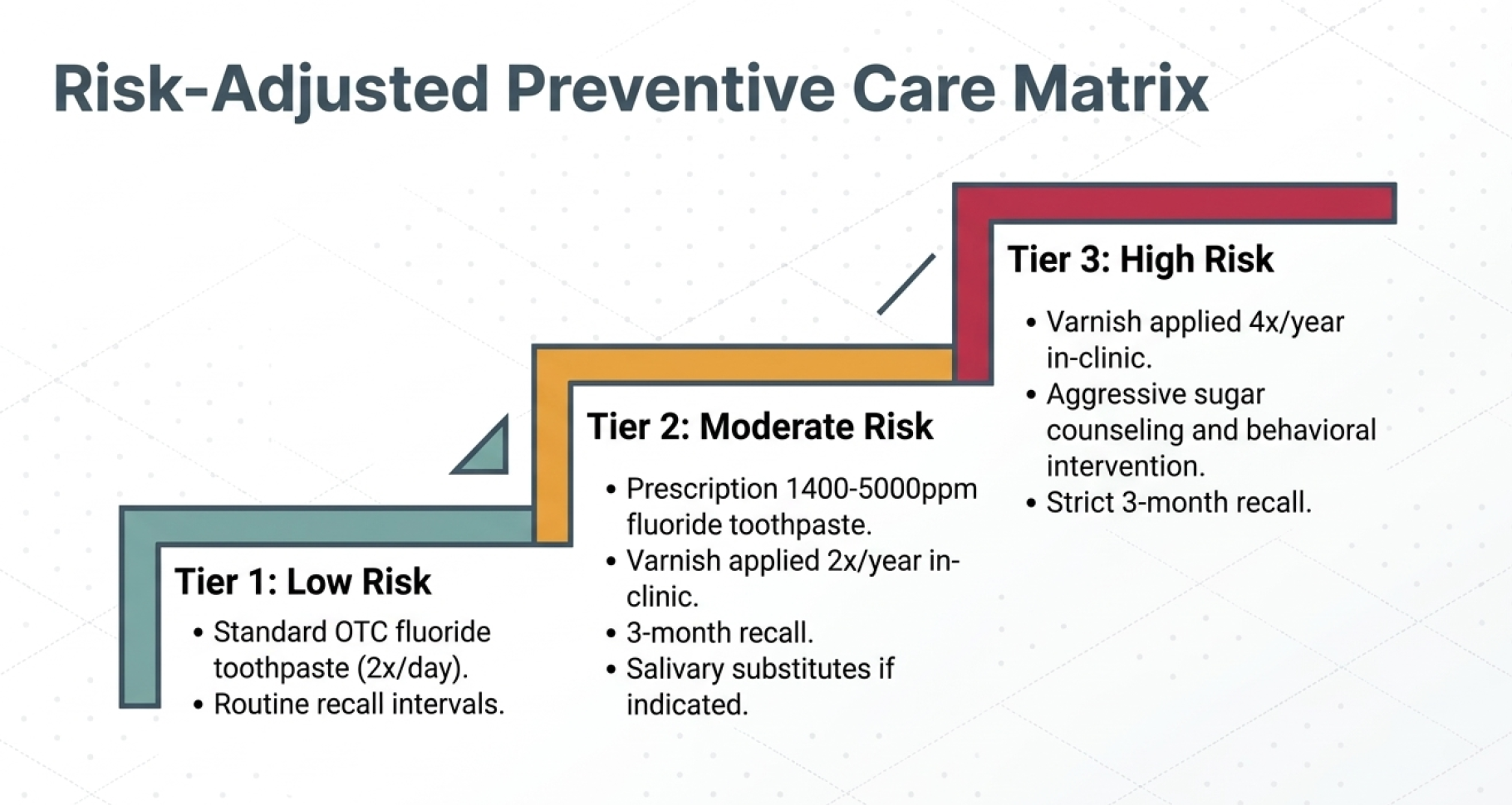
Treatment planning synthesizes the patient's risk category with lesion stage and activity.
1- Non-Operative Care (NOC):
Indicated for Initial active lesions and all inactive lesions. Protocols include high-efficacy fluoridated toothpastes (1450 ppm F⁻), professional topical fluoride varnish applications (2 to 4 times per year based on risk), resin-based or glass ionomer sealants for occlusal pits/fissures, and micro-invasive resin infiltration for non-cavitated proximal lesions.
2- Tooth-Preserving Operative Care (TPOC):
Required for Moderate active and Extensive active lesions. Operative care focuses on selective caries excavation to preserve pulpal vitality, followed by placement of durable direct or indirect restorations.

About the Author:

Dr. Mohamed is a licensed dentist in Canada who successfully passed both AFK and ACJ exams. After seeing too many talented international dentists fail due to poor study strategies, he created AFKStudyPlan to provide structured, evidence-based preparation. He's helped 342+ dentists pass their NDEB equivalency exams.
Have questions?
Email us at Info@afkstudyplan.com
or call 587-707-7068.

Latest from our blog