Sep 27 • AFK study plan

The Ultimate Guide to Oral Pathology: Spotting Tooth Abnormalities, White & Red Lesions, Pigmentation, and Ulcers

Understanding oral pathology is essential for dental professionals, medical practitioners, and board review candidates. From identifying subtle developmental anomalies of the teeth to recognizing potential premalignant oral mucosal lesions, accurate diagnosis begins with clear clinical terminology and thorough visual evaluation. In this comprehensive guide, we unpack five essential domains of oral pathology: basic clinical terminology, tooth shape abnormalities, white and red lesions, oral pigmentations, and mucosal ulcerative conditions.

🎁 Your FREE AFK High-Yield Guide Is Ready!

Get instant access to a high-yield AFK PDF packed with important concepts, key facts, and exam-focused information you need to know.
📘 Study smart. Focus on what matters. Build your confidence.
Good luck with your AFK journey! 🦷💙
Enter your email to get your FREE PDF instantly.
Write your awesome label here.

1. Decoding Basic Oral Pathology Terminology
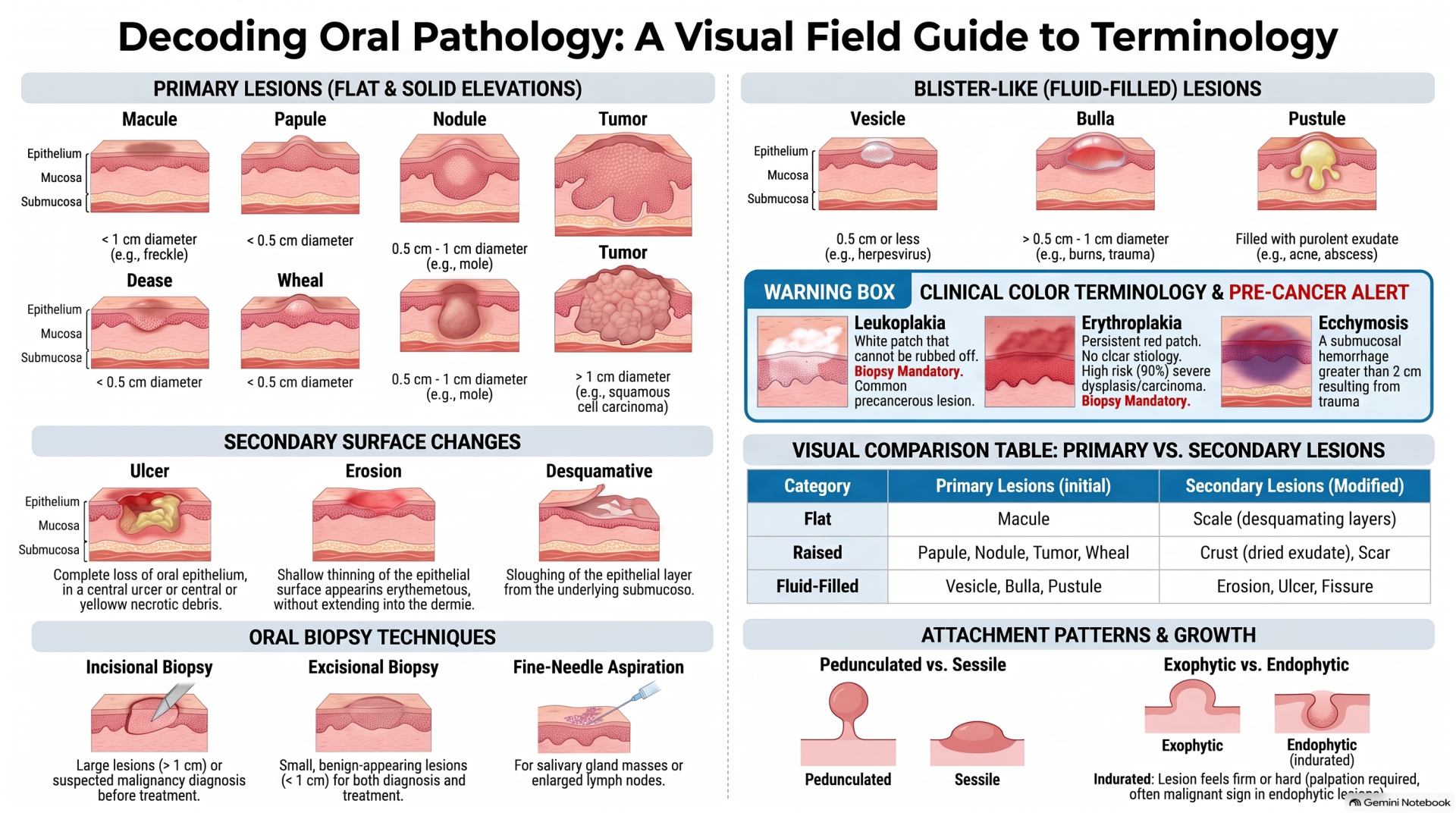
Before diagnosing oral disease, clinicians must master primary and secondary lesion descriptions. Precise
terminology not only aids clinical documentation and medico-legal records but also guides treatment planning and biopsy objectives.
● Macule: A flat, circumscribed area of color change on the oral mucosa with no elevation or depression.
● Papule: A well-circumscribed, solid, raised lesion less than 5 mm (0.5 cm) in diameter.
● Nodule: A solid raised mass larger than 5 mm extending deeper into the tissue.
● Vesicle vs. Bulla: A vesicle is a superficial fluid-filled blister ≤ 5 mm, while a bulla exceeds 5 mm in
diameter containing serum or blood.
● Erosion: Epithelial thinning characterized by an erythematous surface without complete epithelial loss.
● Ulcer: Localized area demonstrating complete loss of the oral epithelium.
● Exophytic vs. Endophytic: Exophytic lesions extend outward from adjacent mucosa, whereas endophytic
lesions extend inward and are typically indurated.
 Clinical Pearl:
 A biopsy is required to establish a definitive histopathological diagnosis, determine prognosis in
premalignant or malignant lesions, and verify treatment efficacy.

2. Tooth Abnormalities: Developmental & Shape Anomalies
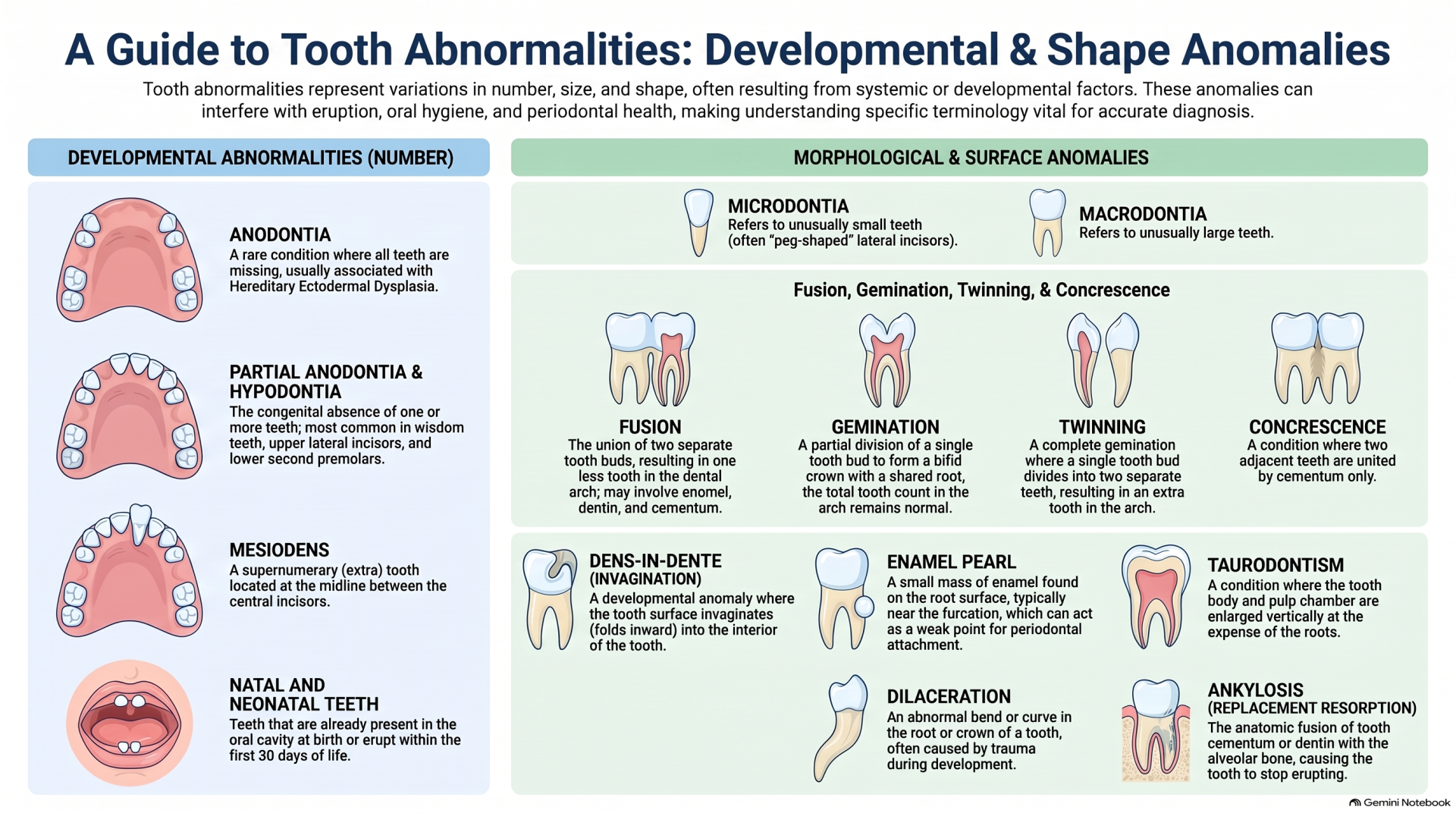
Developmental anomalies of the dentition can affect tooth size, shape, structure, and root morphology.
Shape & Fusion Abnormalities
• Gemination: Partial division of a single tooth bud resulting in a bifid crown sharing one root and pulpal canal. Overall tooth count in the arch remains normal.
• Fusion: Union of two separate tooth buds. Results in a reduced tooth count in the dental arch (minus one).
• Twinning: Complete division of a single tooth bud into two distinct teeth, producing a supernumerary tooth in the arch.
• Concrescence: Union of adjacent teeth along root surfaces by cementum only, most frequently observed in maxillary second and third molars.
• Dens Invaginatus (Dens in Dente): Invagination of enamel organ into the crown before calcification.
Classified as Type I (confined to crown), Type II (extends below CEJ ending in blind sac), and Type III (perforates apically/laterally).
Hereditary Dentin Defects
• Dentinogenesis Imperfecta: Partial Hereditary defect of dentin caused by DSPP gene mutations. Teeth display an opalescent sheen, bulbous crowns, cervical constriction, and rapid pulp obliteration.
• Dentin Dysplasia Type I (Radicular): Features rootless teeth with short/absent roots and spontaneous periapical radiolucencies.
• Dentin Dysplasia Type II (Coronal): Features normal root length, thistle-tube shaped pulp chambers, and frequent pulp stones.

Free Resources to Support Your Preparation 🎥📚🆓

We believe that every candidate should have access to high-quality learning materials, which is why we offer free resources to support your preparation for the AFK exam! 🌟🎯📖

Interactive Videos: Our expertly crafted videos explain key topics in an engaging, easy-to-understand format, helping you grasp essential concepts more effectively.

Question Bank with Detailed Answers: Practice with real exam-style questions and receive comprehensive explanations to strengthen your understanding and improve your problem-solving skills.

One-on-One Consultation: Need a study plan tailored to your unique needs? We offer personalized 1-on-1 sessions, where we assess your situation and learning preferences to recommend the best study strategies for your success. 🤝📈📅

These resources are designed to empower you with the knowledge and confidence needed to excel in the equivalency process. Take advantage of them and set yourself up for success! 🚀📚💡

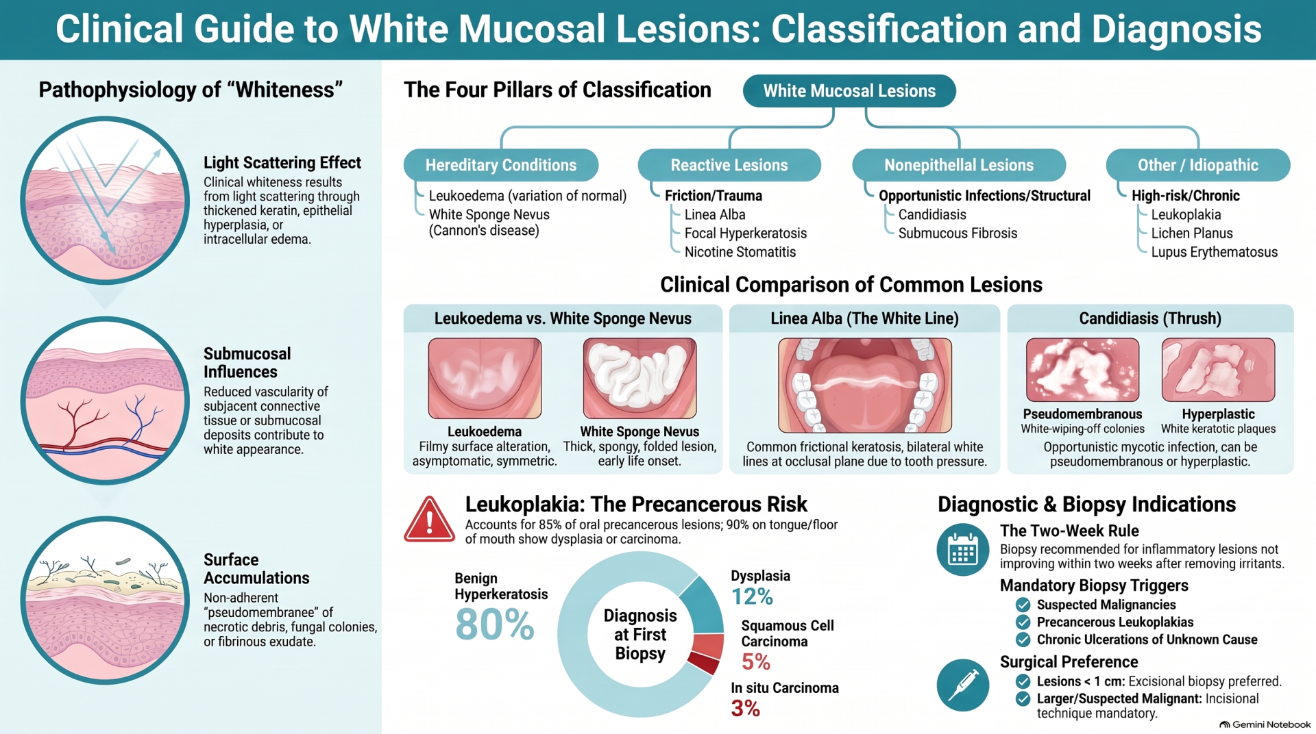
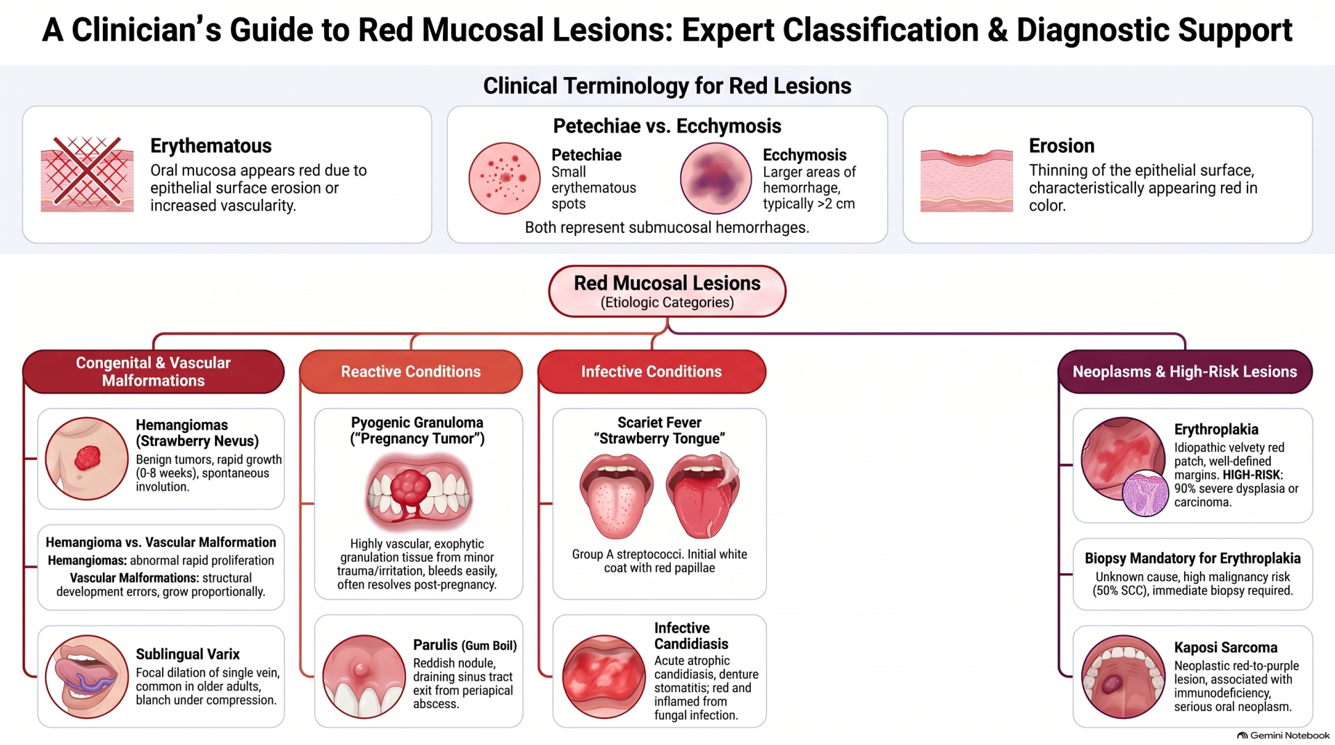
3. White & Red Oral Mucosal Lesions

White and red mucosal lesions range from benign reactive changes to high-risk precancerous conditions.
• Oral Leukoplakia: A clinical white patch that cannot be wiped off or classified as any other entity. Most common precancerous oral lesion (85% of precancers). Lesions on the floor of the mouth and tongue account for over 90% of dysplastic or malignant cases. Mandatory biopsy is required.
• Oral Lichen Planus: Chronic T-cell–mediated (CD8+) autoimmune mucocutaneous disorder featuring Wickham striae (reticular variant) or erythematous/erosive mucosa.
• Oral Candidiasis: Opportunistic fungal infection caused by Candida albicans, presenting in
pseudomembranous (thrush) or erythematous forms.
• Geographic Tongue (Erythema Migrans): Benign shifting erythematous patches with hyperkeratotic
borders caused by desquamation of filiform papillae.

4. Pigmented Oral Lesions: Melanin vs. Exogenous Pigments
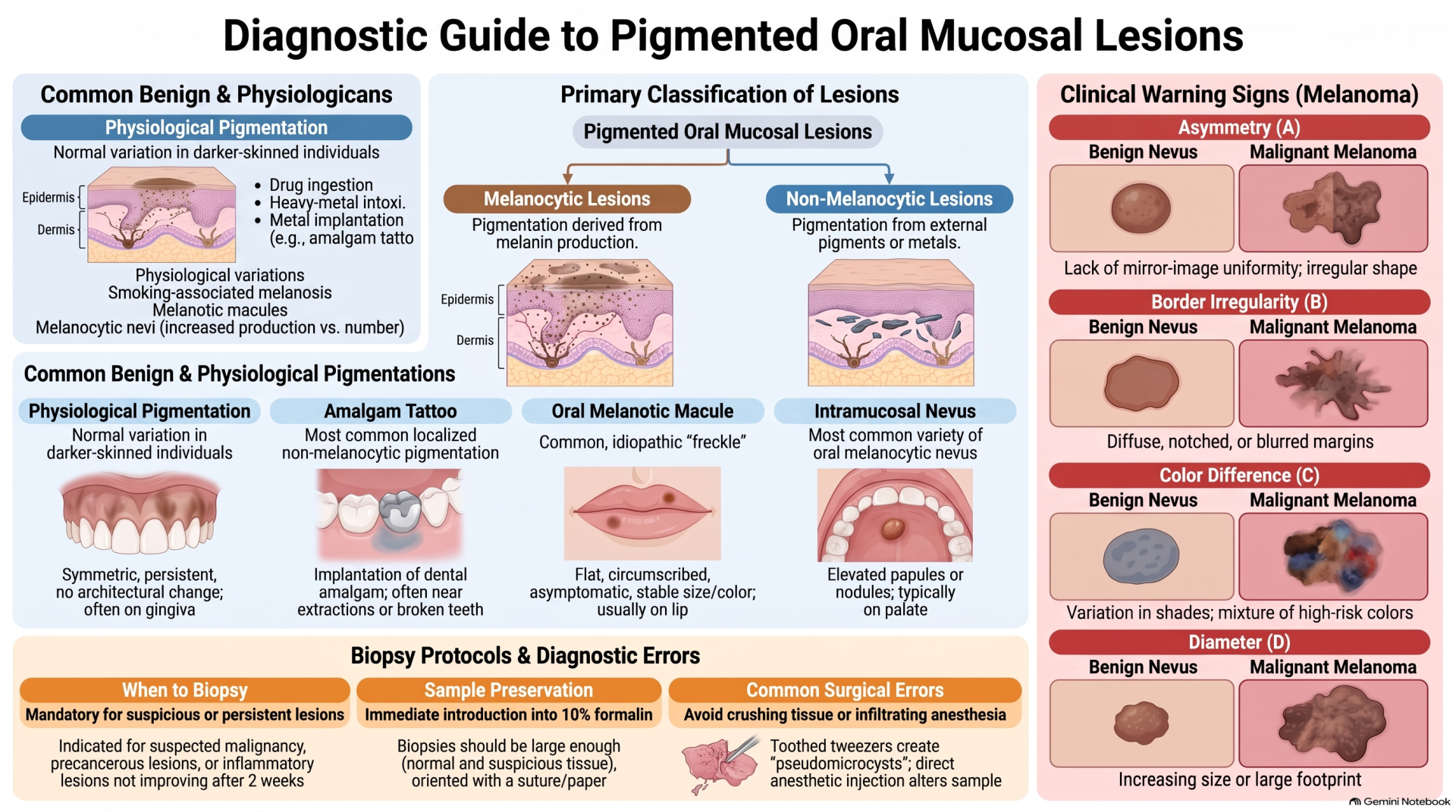
Oral pigmentations are classified as melanocytic (melanin-derived) or non-melanocytic (exogenous deposits
like metals or amalgam).
• Amalgam Tattoo (Focal Argyrosis):
Most common oral mucosal pigmentation. Iatrogenic lesion caused by
traumatic soft-tissue implantation of amalgam particles. Appears as a flat, gray-blue macule.
• ABCD Criteria for Oral Melanoma: Evaluates Asymmetry, Border irregularity, Color variation, and
Diameter growth for suspicious melanocytic lesions.
• Heavy Metal Lines: Ingestion of heavy metals like bismuth or lead can deposit along the gingival margin as
blue-black linear discolorations (bismuth or lead lines).

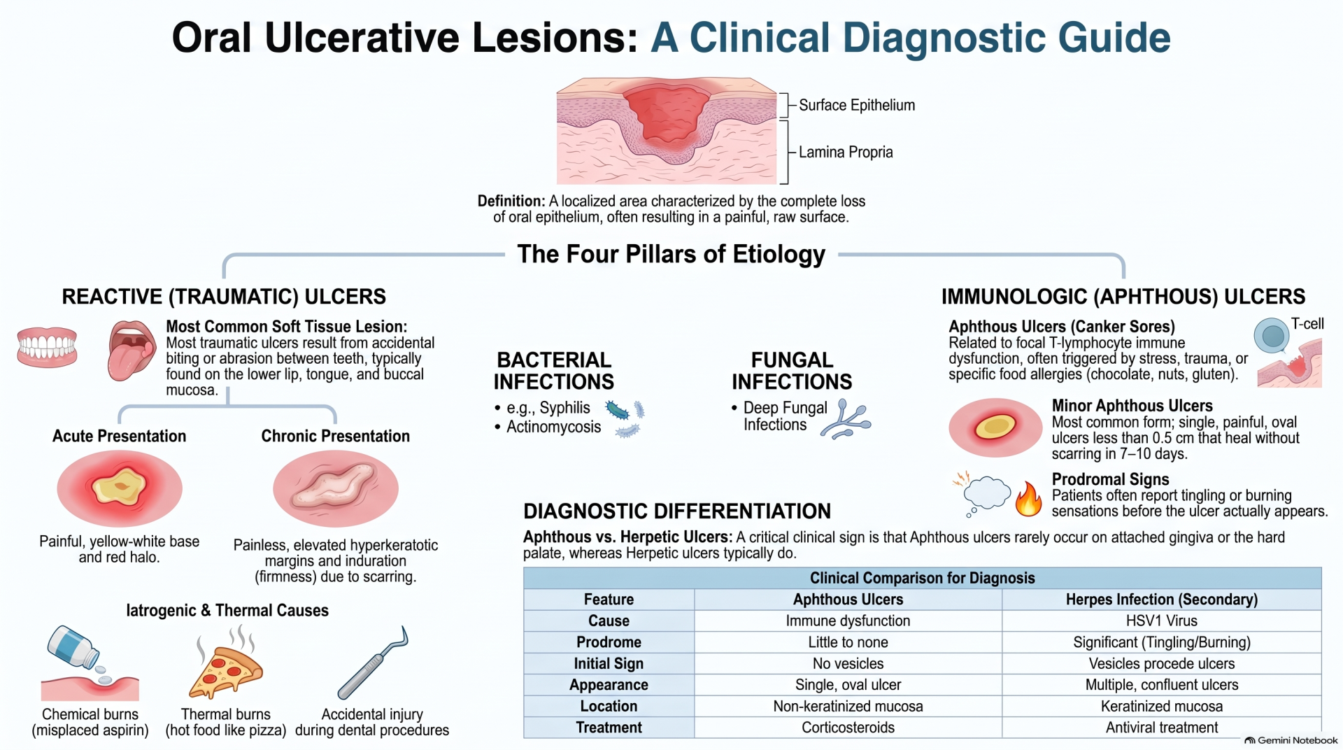
5. Mucosal Ulcerative Diseases

Oral ulcers result from complete loss of surface epithelium, stemming from reactive trauma, infections,
immunologic disorders, or neoplasms.
• Traumatic Ulcers: Most common oral soft-tissue lesions. Acute ulcers present with a yellow-white fibrinous
base and erythematous halo. Chronic ulcers feature raised, hyperkeratotic borders.
• Recurrent Aphthous Stomatitis (Canker Sores): Painful, recurrent oval ulcers surrounded by an
erythematous halo occurring almost exclusively on non-keratinized mucosa.
• Infectious & Immunologic Ulcers: Includes viral infections (HSV, VZV), autoimmune blistering diseases
(Pemphigus, Pemphigoid), and malignant ulcerations (Squamous Cell Carcinoma).

About the Author:

Dr. Mohamed is a licensed dentist in Canada who successfully passed both AFK and ACJ exams. After seeing too many talented international dentists fail due to poor study strategies, he created AFKStudyPlan to provide structured, evidence-based preparation. He's helped 342+ dentists pass their NDEB equivalency exams.
Have questions?
Email us at Info@afkstudyplan.com
or call 587-707-7068.

Latest from our blog